Project is in private repo due to Massachusetts General Hospital/Harvard Medical School intellectual property regulation.
Introduction
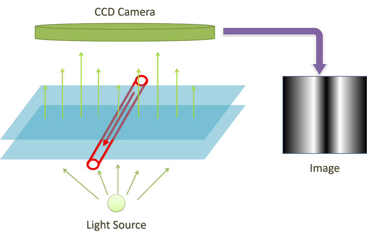
The Project aim to develop a functional system to image the blood vessels in a non-invasive manner, and also able to distinguish between artery and vein. According to diffuse optical principles, oxygenated and deoxygenated blood and water have very different extinction coefficients. Here we use Near Infrared Light of two wavelengths (690nm | 830nm) as the light source. And then apply adaptive Frangi-Hessian vesselness filter to extract the linear structures and enhance the image with Contrast Limited Adaptive Histogram Equalization (CLAHE) to obtain the image frame for visualization.
Vascular Structure Enhancement
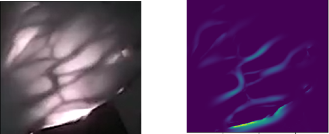
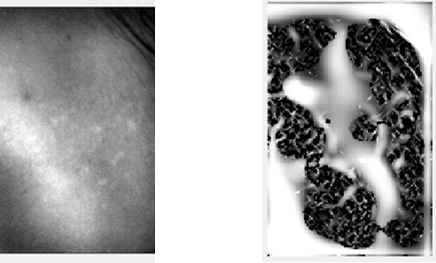
In Frangi-Hessian filtering, best performance is achieved when the sigma parameter is set similar to the scale of the linear structure. And beta parameter determines the Tolerance of the algorithm to detect inflection points. Here are the results when Beta is set to 4, sigma is iterates from 1-to-10 (pixels).
- Imaging the Hand
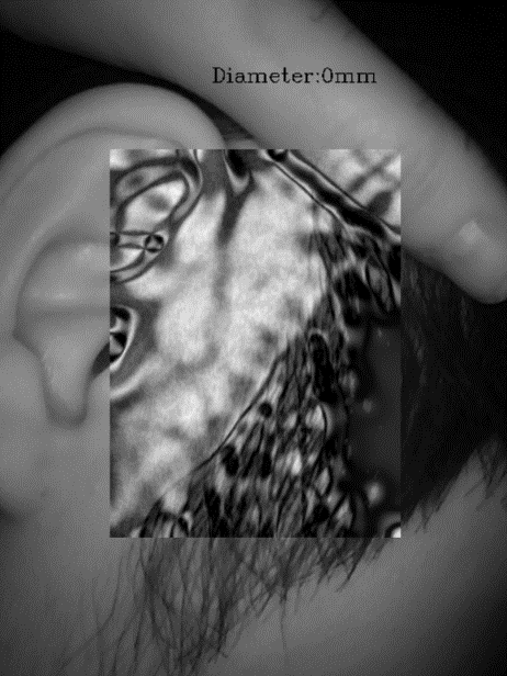
- Imaging the superficial Temporal Artery(STA)
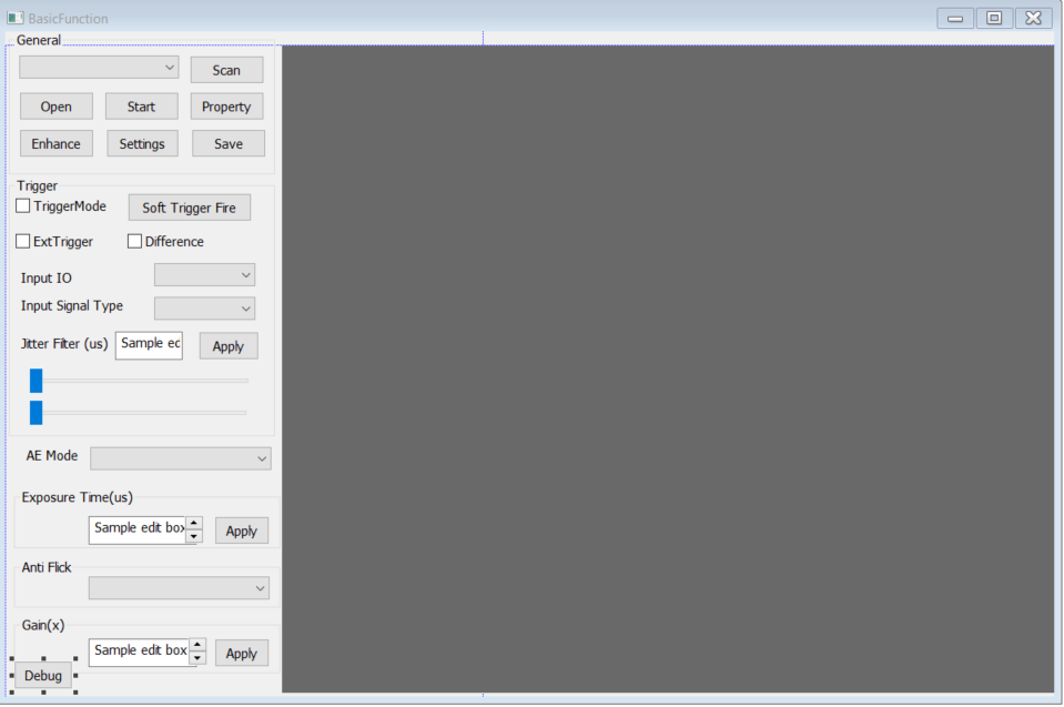
The Visualization APP
The visualization is implemented with DoThink3 camera sdk and MFC. Built in anti-flicker, parameter adjusting scroll bar, external trigger mode, auto exposure and many others.
Human Subject Trial
Graduate Thesis
©Massachusetts General Hospital/Harvard Medical School reserves the rights associated with the innovations and intellectual properties in this thesis work.